Okayama University Medical Research Updates (OU-MRU) Vol.3
November 30, 2014
Source: Okayama University (JAPAN), Center for Public Information
For immediate release: 30 November 2014
Okayama University research: Organ regeneration research leaps forward
(Okayama, 30 November 2014) Researchers at Okayama University Graduate School of Medicine and Kyorin University School of Medicine have successfully generated a kidney-like structure from just a single cell.
“It has been predicted that the kidney will be among the last organs successfully regenerated in vitro due to its complex structure and multiple functions,” state Shinji Kitamura, Hiroyuki Sakurai and Hirofumi Makino at the beginning of their latest report, before continuing to describe results suggesting a far more positive prognosis for the pace of kidney regeneration research. Despite the anatomical challenges posed by the kidney anatomy and the complexities understood from embryonic kidney development processes, the researchers have demonstrated that kidney-like structures can be generated from just a single adult kidney stem cell.
In embryos, kidney development requires two types of ‘primordial’ cells – cells at the earliest stage of development. However by generating kidney-like structures from a single type of kidney stem cell the researchers provide evidence for differences in the organ development in adults and embryos.
Kitamura, Sakurai and Makino – researchers from Okayama and Kyorin Universities - took kidney stem cells from the different kidney components of microdissected adult rats and grew them in culture. A method for growing three-dimensional cell clusters showed that kidney-like structures could form so long as the initial cell cluster was large enough.
The minimum cluster size required might suggest that not all the kidney stem cells have stem cell characteristics. Therefore the researchers cloned kidney stem cells and confirmed that kidney-like structures still formed from the clusters of clone cells after a few weeks.
The researchers add, “Although the physiological roles of such cells are currently unclear, analogous cells in the adult human kidney would be a valuable resource for the regeneration of kidneys in vitro.”
Background
Kidney structure
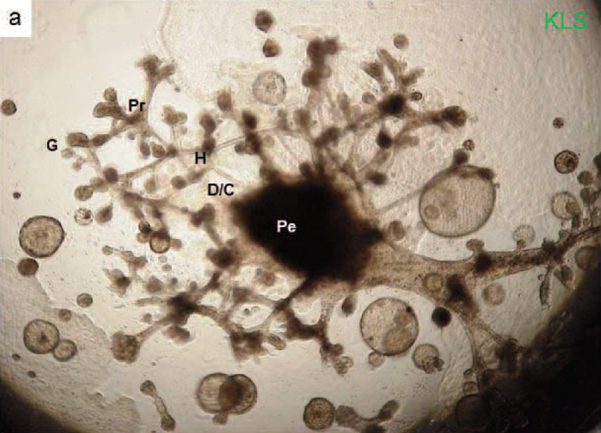
There are more than a dozen distinct types of cell in the kidneys. The basic structural unit of the kidney is the nephron, which filters the blood to regulate the concentration of water and soluble substances such as sodium salts. Each nephron comprises several well-defined segments: the glomerulus, the proximal tubule, the loop of Henle, the distal tube and the collecting duct.In embryo kidney organogenesis two primordial cell types are required to differentiate into all the different cell types in the kidney: metanephric mesenchymal cells and uteric bud cells. Kitamura, Sakurai and Makino produced kidney cells that could differentiate into a kidney-like structure without these primordial cell types, suggesting these are adult kidney stem cells.
Obtaining kidney stem cells
The researchers microdissected adult rat kidneys into segments from the glomeruli, proximal convoluted tubule (S1/PCT), proximal straight tubule (S2, S3), medullary thick ascending limb of Henle’s loop and the collecting duct. They then grew the cells on mouse mesenchymal cells. While there is no known single biomarker for adult kidney stem cells, immunohistochemical anaylysis identified a number of markers in the kidney stem cells- that are found in embryonic or adult kidneys.Three-dimensional culture and morphogenesis
The cells were initially grown on type IV collagen and in two-dimensional growth conditions there were no signs of kidney structure development. Instead the researchers used a ‘hanging drop’ method for making cell cluster to generate 3-dimensional structure. Cell clusters were placed in a half MatrigelTM - a gelatinous protein mixture secreted by Engelbreth-Holm-Swarm mouse sarcoma cells. This protein gel provides a complex extracellular environment similar to that found in many tissues.The cell clusters cultured in this way reproducibly formed kidney-like structures depending on the size of the initial cell cluster. When less than 6250 cells were used only cystic structures developed. When the initial cluster contained up to 50,000 cells distinct tubular structures were observed and when the cluster size exceeded 100,000 cells full kidney-like structures containing a glomerulus, as well as proximal and distal tubules developed.
The kidney-like structures formed did not show the formation of blood vessels or make urine. Further research is required to understand these aspects of kidney regeneration.
Acknowledgments
A portion of this study was supported by the Health and Labor Sciences Research Grants for Research on Tissue Engineering from the Ministry of Health Labor and Welfare of Japan, the programme for the promotion of fundamental studies in Health Sciences of the National Institute of Biomedical Innovation (VIBIO), a Grant-in-Aid for Young Scientists (B) and a grant from the Okayama Medical Foundation, the Ministry of Economy, Trade and Industry (METI), Japan, and joint research with Organ Technologies, Inc. We appreciate the technical contributions of Dr. Takeshi Sugaya (Cimic, Co., Ltd., Japan) and Dr. Naoshi Shinozaki (Cornea Center & Eye Bank, Tokyo Dental College, Ichikawa General Hospital) and the kind advice of Dr. Kazuhiro Sakurada (Sony Computer Science Laboratories, Inc., Japan) and Dr. Naoya Kobayashi (Okayama Saidaiji Hospital).Reference
Shinji Kitamura, Hiroyuki Sakurai, Hirofumi Makino, “Single adult kidney stem/progenitor cells reconstitute three-dimensional nephron structures in vitro” , Stem Cells, x, (x), pp.yy-zz, (2014) in press.Abstract URL: http://onlinelibrary.wiley.com/doi/10.1002/stem.1891/abstract
DOI: 10.1002/stem.1891

Figure caption:
The relationship between cell number in the cluster and the ability to reconstitute a kidney-like structure. Red bar: cyst formation, Blue bar: long distinct tubules formation, Green bar: tubules with ball-like structures at the tip. The horizontal axis represents kidney stem/progenitor cell (KS cell) number (6.25–200 × 103 cells)/cluster. The KS cell clusters were cultured for 3 weeks. Representative photomicrographs were from three independent experiments. Scale bar = 100 μm.

Figure caption:
A KS cell cluster formed a kidney-like structure (KLS) after 4weeks of incubation using DMEM/F12 plus 10% FCS, 250ng/ml GDNF, 250ng/ml b-FGF, 250ng/ml HGF, 250ng/dl BMP-7 and 500 ng/ml EGF. Glomerulus-like structures were formed at the tips of the tubular structures, proximal like tubules, distal like tubules, collecting duct-like tubules and renal pelvis-like structures. G, glomerulus-like structure; Pr, proximal tubule-like structure; H, loop of Henle-like structure; D/C, distal tubule-like structure or collecting duct-like structure; Pe, renal pelvis-like structure.
Correspondence to
Senior Assistant Professor Shinji Kitamura, M.D., Ph.D.Department of Medicine and Clinical Sciences, Okayama
University Graduate School of Medicine, Dentistry and
Pharmaceutical Sciences, 2-5-1 Shikata-cho, Kita-ku,
Okayama 700-8558, Japan
E-mail: [email protected]
Further information
Okayama University1-1-1 Tsushima-naka , Kita-ku , Okayama 700-8530, Japan
Planning and Public Information Division, Okayama University
E-mail: [email protected]
Website: //www.okayama-u.ac.jp/index_e.html
Okayama University Medical Research Updates (OU-MRU)
Vol.1:Innovative non-invasive ‘liquid biopsy’ method to capture circulating tumor cells from blood samples for genetic testingVol.2:Ensuring a cool recovery from cardiac arrest
About Okayama University
Okayama University is one of the largestcomprehensive universities in Japan with roots going back to the Medical Training Place sponsored by the Lord of Okayama and established in 1870. Now with 1,300 faculty and 14,000 students, the University offers courses in specialties ranging from medicine and pharmacy to humanities and physical sciences. Okayama University is located in the heart of Japan approximately 3 hours west of Tokyo by Shinkansen.